
MAP YOUR PAIN
Take the Free Pain Survey
Occipital Nerve Decompression Surgery (NDS)
Written and medically reviewed by Carlton Perry, MD, board-certified by the American Board of Plastic Surgery · Last reviewed
If you have severe head or neck pain, have tried treatment after treatment, and still hurt every day, it is reasonable to wonder whether something else should be considered.
For some patients, the missing piece is an injured, irritated, or compressed peripheral nerve. Occipital nerve decompression is a focused operation that releases pressure around the affected nerves. The goal is to preserve the nerves, improve their local environment and circulation, and give them a better opportunity to recover—not intentionally destroy them.
There is no implanted battery, pulse generator, or permanent stimulation system. This is a hardware-free operation, but it is not simply a hardware-free version of nerve stimulation. Decompression and stimulation are different treatments with different indications, risks, and reasons they may or may not succeed.
Most importantly, this operation is not appropriate for every person with chronic headache. The operation matters, but choosing the right patient and the right surgeon may matter just as much.
Show us where your pain is—where it starts and where it goes. Our Free Pain Survey includes an interactive 3D model that can help you describe a pain pattern that may be difficult to put into words.
What is occipital nerve decompression?
The greater, lesser, and third occipital nerves carry sensation from the upper neck and scalp. Along their paths, they travel through muscle and fascia and may cross or run beside blood vessels. Trauma, previous surgery, scarring, inflammation, or anatomic compression can sometimes make one or more of these nerves a persistent source of pain.
During surgery, Dr. Perry identifies the involved nerves and follows them through areas where they may be tethered or compressed. Tight fascia, scar tissue, portions of muscle, or a closely associated vessel may be addressed when indicated.
These are sensory nerves that supply the scalp; they do not control movement. The operation takes place in the soft tissues of the upper neck and scalp. It is not brain surgery, does not enter the skull, and is not performed in the spinal cord.

Why patient selection matters
Two people can carry the same diagnosis and still have very different pain generators. A diagnosis such as occipital neuralgia does not, by itself, establish that surgery is appropriate.
The evaluation brings together the patient’s history, pain map, physical examination, anatomy, and response to previous treatments. Before recommending surgery, the surgeon should have reasonable confidence that the patient’s pain includes a meaningful occipital nerve component that may be addressed through decompression.
Published surgical studies generally involve carefully selected patients whose symptoms persisted despite substantial nonsurgical treatment. Even in those selected groups, improvement varies. The purpose of screening is not to find a reason to operate on everyone. It is to determine whether the history and pain pattern justify closer evaluation of a peripheral nerve source.
How the involved nerves are identified
The process begins with the pain pattern: Where does the pain start, and where does it go? Pain that begins in the head or neck and travels across the scalp can tell a different story from pain that begins at the forehead, temple, or behind the eye, although both patterns can sometimes include an occipital nerve component.
The pain map is compared with focal tenderness, trigger points, scalp sensitivity, numbness, tingling, and reproduction of the patient’s familiar pain during examination. Previous responses to nerve blocks, injections, Botox, radiofrequency treatment, surgery, or nerve stimulation may add useful information. Imaging can help exclude other conditions, but the decision is rarely made from an MRI or any other single test.
Show us where your pain is—where it starts and where it goes. Start the Free Pain Survey
What happens during surgery?
Dr. Perry performs occipital nerve decompression under general anesthesia. The operative evaluation includes exploration of the midline and both sides of the posterior head and neck. Nerve decompressions are performed where indicated.
The operation releases structures that are constricting, tethering, or irritating the involved nerves. Depending on the patient’s anatomy, this may include fascia, muscle, scar tissue, or a vessel closely crossing or intertwining with a nerve. When decompression is the appropriate procedure, the nerve is kept intact. A severely injured nerve or painful neuroma may require a different nerve-directed operation.
The operation is performed as an outpatient procedure, and patients ordinarily go home the same day. Rarely, additional observation may be necessary because of anesthesia recovery, nausea, an unexpected medical concern, or another individual circumstance. Every patient receives detailed postoperative instructions.
How we think about results
Published studies report meaningful improvement in carefully selected patients with persistent occipital neuralgia and related peripheral nerve pain patterns. There is no single universal success rate because studies use different diagnoses, selection criteria, procedures, follow-up periods, and definitions of success.
We follow changes in the patient’s overall Headache Burden—not just one pain score. Improvement may mean fewer pain days, lower pain intensity, shorter severe episodes, less medication, better sleep, or the ability to return to work and activities.
One of the most useful questions is: What would you like to do after surgery that you cannot do now because of your head and neck pain?
Surgery may substantially reduce the nerve-generated component of pain, but it cannot guarantee complete pain freedom or eliminate a separate headache disorder.
The operation matters. Patient selection matters just as much.
Occipital nerve decompression is not appropriate for every person with chronic headache. A diagnosis alone does not establish that surgery is the right next step.
The evaluation brings together the pain pattern, examination, anatomy, and response to previous treatments. Before recommending surgery, the surgeon should have reasonable confidence that the pain includes a meaningful occipital nerve component.
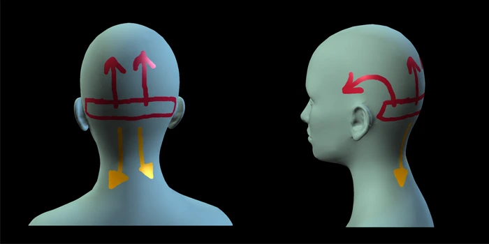
Pain location and direction can provide important clues. The Free Pain Survey allows you to show us where your pain starts and where it goes using an interactive 3D model. Illustrative map created with fictional patient information. Everyone’s pain map is unique.
Frequently Asked Questions
1. Who may be a candidate for occipital nerve decompression surgery?
A potentially appropriate candidate usually has a coherent peripheral nerve pattern rather than simply severe or frequent headaches. Helpful findings may include pain beginning in the distribution of a specific nerve, focal tenderness or a trigger point over that nerve, scalp sensitivity or altered sensation, a history consistent with nerve injury or compression, and symptoms that have not improved adequately with reasonable nonsurgical care.
A targeted nerve block or another treatment that changes the characteristic pain can add useful information, but no single test makes the decision by itself. Good candidacy also requires realistic goals. The purpose is to reduce the part of the pain being generated by the involved nerves, not promise that every headache will disappear permanently.
2. What makes someone a poor candidate?
Surgery becomes less compelling when the pain has no consistent anatomic pattern, there is no focal tenderness or sensory change, targeted diagnostic treatment repeatedly has no effect, or another untreated condition better explains the symptoms.
Other concerns include active infection, medical problems that make elective surgery unsafe, inability to follow postoperative instructions, or expectations that no operation can realistically meet. A careful “not yet” or “this is not the right operation” can be as important as recommending surgery.
Sources: [1]
3. Do I have to try every possible treatment before considering surgery?
No. Patients should not have to collect failed treatments simply to earn an operation. At the same time, decompression is usually considered after appropriate nonsurgical care has failed, stopped helping, or provided only temporary relief.
The relevant treatment history differs from patient to patient. It may include medications, targeted injections or nerve blocks, Botox, physical therapy, or other treatments chosen for the diagnosis. What matters is the collective pattern of symptoms, clinical findings, and treatment responses—not an arbitrary number of failed therapies.
Sources: [3]
4. How does the surgeon decide which nerve is involved?
The decision is based on pattern recognition across the history, 3D pain map, physical examination, anatomy, and previous treatment responses. Patients can create their own 3D pain map in the Free Pain Survey, showing where the pain starts, where it travels, and how it moves. A targeted nerve block that temporarily changes one recognizable part of the pain may be informative. Previous trauma, surgery, radiofrequency treatment, Botox, or nerve stimulation can also affect the anatomy and interpretation of symptoms.
Before recommending surgery, the surgeon should be able to explain why the patient’s symptoms appear to include a meaningful occipital nerve component and which nerves may need to be addressed.
5. What happens during occipital nerve decompression surgery?
The basic goal is straightforward: identify the painful occipital nerve, follow it through the areas where it can be constricted, and release the structures that are compressing or tethering it. Depending on the nerve and the patient’s anatomy, those structures may include tight fascia, portions of muscle, scar tissue, or a vessel that is intimately crossing or intertwining with the nerve.
The exact technique varies among surgeons. Published approaches include midline posterior incisions, separate or combined approaches to the lesser occipital nerve, and endoscopic-assisted techniques. The nerve is generally preserved when decompression is the goal. Revision cases, neuromas, or severely injured branches can require a different strategy, such as neurectomy with management of the nerve end.
This is not brain surgery and it does not involve opening the skull. It is peripheral nerve surgery in the soft tissues of the scalp and upper neck. The operative plan should specify which nerves will be treated and whether any prior scars, stimulator leads, or nerve injuries change the approach.
6. How are the greater, lesser, and third occipital nerves decompressed?
The greater occipital nerve travels through several possible compression zones in the upper neck before reaching the scalp. Adequate decompression may require releasing it along the relevant part of that course rather than opening only one small point.
The lesser occipital nerve travels farther to the side and has different potential entrapment points. The third occipital nerve is smaller and is addressed when the pain map, examination, and operative findings support its involvement.
Anatomic variation is very common. Branching patterns, vessel crossings, and connections between nerves frequently differ from medical illustrations. Experience matters because the surgeon must recognize both expected anatomy and the variations encountered during an operation.
7. Can an occipital nerve recover after years of compression?
Sensory nerves may retain the ability to recover even after prolonged irritation or compression. This differs from recovery in a motor nerve, where prolonged loss of nerve supply can lead to irreversible changes in the muscle.
Recovery still depends on the severity and duration of the injury, whether the nerve remains intact, previous procedures, scar tissue, and individual healing. Decompression creates a better environment for recovery, but it cannot guarantee complete pain relief or the return of normal sensation.
8. What does blood flow have to do with nerve decompression?
When an occipital nerve is compressed, its local circulation may also be affected. During surgery, Dr. Perry often sees a visible change in the nerve after constricting tissue is released, including improvement in its color and appearance.
Operative photographs are taken to document each patient’s findings and are reviewed with the patient after surgery. Decompression relieves mechanical pressure around the nerve and may improve its local circulation, creating a better environment for recovery. The photographs document visible operative findings; they do not directly measure microscopic blood flow within the nerve.
9. Are frontal or temporal nerves ever decompressed?
The supraorbital, supratrochlear, and auriculotemporal nerves can be peripheral pain generators, but they are treated far less often in the patient population discussed here.
Pain in the forehead, temple, or behind the eye does not automatically mean that a frontal or temporal nerve needs surgery. Occipital pain can travel forward. If the pain consistently starts in the back of the head and then reaches the eye or temple, treating the occipital source may make more sense than adding an unnecessary frontal or temporal procedure.
Sources: [11]
10. Is occipital nerve decompression minimally invasive, and how serious is it?
Occipital nerve decompression is less invasive than an operation that enters the skull or cervical spine, but it is still real surgery. It requires an incision, careful nerve dissection, release of constricting tissue, and general anesthesia. Calling it minor surgery can give patients the wrong expectation.
“Focused peripheral nerve surgery” is a more accurate description. It is anatomically very different from a craniotomy, cervical fusion, or permanent neurostimulation implant.
11. Is general anesthesia required, and can I go home the same day?
Dr. Perry performs occipital nerve decompression under general anesthesia. It is an outpatient procedure, and patients ordinarily go home the same day.
Rarely, additional observation may be necessary because of anesthesia recovery, nausea, an unexpected medical concern, or another individual circumstance. Every patient receives detailed instructions covering medications, fasting, transportation, postoperative supervision, and when to contact the surgical team.
12. Does age determine whether someone can have surgery?
Age alone does not determine candidacy. The more important questions are whether the diagnosis is sound, whether peripheral nerves are truly involved, whether appropriate nonsurgical care has been tried, and whether the patient can safely undergo surgery.
In younger patients, evolving headache disorders and other causes require particular care, and the published evidence is stronger in adults than in children. In older adults, medical conditions, anticoagulant use, wound healing, cervical spine disease, and other causes of new headache become increasingly important. There is no absolute rule that someone is automatically too young or too old.
13. Can I have nerve decompression if I have had prior head, neck, brain, or cervical spine surgery?
Possibly. Prior surgery can actually be part of the reason a peripheral nerve is painful. Craniotomy, cervical spine procedures, scalp surgery, and other operations can injure an occipital nerve directly or create scar around it. A 2024 series of iatrogenic occipital nerve injuries reported substantial improvement after decompression or neuroma-directed surgery in carefully selected patients.
Previous surgery also makes the operation more complex. Scar changes the normal tissue planes, the nerve may have been cut or stretched, and a neuroma may need a different treatment from simple decompression. Operative reports and old imaging can be extremely valuable.
A history of cervical spine disease does not automatically exclude decompression, but it raises the possibility that more than one pain generator is present. The goal is to identify which part of the pain is realistically addressable by peripheral nerve surgery.
14. How painful is the recovery after nerve decompression surgery?
Most patients have incisional soreness, tightness, swelling, and a different kind of discomfort from their preoperative nerve pain. The first several days are usually the most uncomfortable. Some people notice early relief of their familiar pain; others have temporary numbness, hypersensitivity, zaps, or burning as the nerve settles.
Recovery should not be judged by the first week alone. A nerve that has been chronically irritated can remain reactive after it is released, and the surrounding soft tissues also need time to heal. Postoperative pain control is individualized and may include non-opioid medication, short-term prescription analgesics, local anesthetic strategies, ice or other measures, and instructions tailored to the surgeon’s protocol.
If pain is rapidly worsening rather than gradually stabilizing, or is accompanied by fever, spreading redness, drainage, new weakness, or another concerning symptom, the surgical team should be contacted promptly.
Sources: [6]
15. How soon after surgery will I know whether it worked?
Some patients know very early that the old pain is different. Others do not. Early swelling, bruising, surgical soreness, temporary numbness, and nerve irritability can make the first few weeks noisy from a diagnostic standpoint.
I generally separate “early signs of improvement” from “final result.” If the specific preoperative trigger point is less tender, the familiar shooting pain has changed, or a patient can lie on the back of the head more comfortably, those can be encouraging early signs. But nerves heal slowly, and symptoms can fluctuate as activity increases.
Published studies commonly report formal outcomes at months rather than days. That is a better way to think about the process. The operation happens in one day; the biologic recovery of an injured nerve and the nervous system’s adaptation to a quieter pain signal take longer.
16. How long does it take to see the full result after headache nerve decompression?
There is no single deadline. Soft-tissue healing occurs over weeks, while nerve recovery and remodeling can continue for months. Patients with very long-standing pain may also have central sensitization — the nervous system has learned and amplified the pain signal over years — so the clinical improvement may lag behind the mechanical decompression.
This is why I am reluctant to declare success or failure too early. Published studies often use 12-month outcomes, and long-term series have followed patients for several years. A patient may improve quickly, gradually, in steps, or with temporary setbacks during healing.
The most useful follow-up measures are the same ones we cared about before surgery: number of pain days, average intensity, duration of severe episodes, medication use, sleep, ability to work, and return to activities. Those functional changes are often more meaningful than asking whether every sensation is gone.
17. When can I return to work after nerve decompression surgery?
That depends much more on the type of work than on the job title. Someone working from home at a computer may be able to resume limited work relatively early. A job that involves heavy lifting, repeated neck motion, overhead work, protective headgear, or risk of impact usually requires more time.
Fatigue is also real. Even when the incision looks good, anesthesia, disrupted sleep, postoperative medication, and nerve irritability can make a full workday difficult at first. I would rather have a patient return in a staged fashion than feel pressured to prove the operation was “easy.”
This answer should ultimately be customized to the specific operative technique used by the practice. For the website, it is safest to give patients a general expectation and then make clear that their surgeon will provide the actual work release based on their procedure and occupation.
Sources: [6]
18. What activities should I avoid while I am healing?
Early restrictions usually focus on protecting the incision and preventing strain or trauma to the surgical area. Heavy lifting, vigorous upper-body exercise, repetitive neck exertion, contact sports, and activities that put direct pressure on the back of the head may be limited for a period of time.
Walking and gentle movement are often encouraged sooner than strenuous exercise, but the exact progression is surgeon-specific. The number of nerves treated, whether prior scar tissue or stimulator hardware was involved, and how the wound is healing can all change the timeline.
The right question is not “When can I exercise?” but “When can I safely return to this specific activity?” A stationary bike, lap swimming, weight training, tennis, and a contact sport place very different stresses on the healing tissues.
Sources: [6]
19. What medications will I need after surgery?
There is no universal medication list. Postoperative prescriptions are based on the operation, allergies, other medical conditions, bleeding risk, prior medication use, and the surgeon’s protocol. Pain control may involve acetaminophen, anti-inflammatory medication when appropriate, a short course of stronger analgesia, medication for nausea, or other targeted treatments.
Patients who have been on long-term headache preventives, anticonvulsants, antidepressants, or opioids should not abruptly stop them unless the prescribing physician gives a plan. One of the goals after a successful operation may be to reduce medication burden, but that is usually done gradually and in coordination with the clinicians who manage those drugs.
For the website version, the safest message is that postoperative medication is individualized and that patients should follow the written surgical instructions rather than copying another patient’s regimen.
Sources: [2]
20. Will the scars be visible?
Occipital decompression incisions are generally placed within or near the hair-bearing scalp or upper neck, depending on the technique. Frontal or temporal approaches, when they are truly needed, use different incision locations. Scar visibility depends on the incision, hair pattern, healing tendency, prior surgery, skin type, and whether a patient develops a widened or hypertrophic scar.
The practical tradeoff is that the incision has to be long enough to safely identify and release the nerve. A tiny incision is not automatically better if it limits visualization and leaves compression untreated. The surgeon should be able to show you where the planned incisions will be before the operation.
For patients with prior stimulator surgery or other scars, the old incisions may influence the new approach. Those cases deserve an individualized discussion rather than a generic promise that scars will be invisible.
21. What are the risks and possible complications?
Every operation has risk. Possible problems include bleeding or hematoma, infection, wound-healing difficulty, temporary or persistent numbness, hypersensitivity, painful scar, incomplete relief, recurrence, nerve injury, neuroma, altered or uneven sensation, and the possibility of revision surgery. General anesthesia carries its own risks.
The risk profile changes depending on the nerves treated and whether this is a first operation or a revision through previous scar. A responsible discussion should include common temporary symptoms, less common serious complications, what can be done if a problem occurs, and the surgeon’s own experience with revision.
22. How is improvement measured after surgery?
We look at the patient’s overall Headache Burden. That can include the number of pain days, average intensity, duration of severe episodes, medication use, sleep, work, and the ability to return to meaningful activities.
A lower pain score matters, but it is not the only measure of success. The practical question is what the patient can do after surgery that pain prevented before surgery.
23. Can the pain come back after decompression surgery?
Yes. Durable relief is possible, but recurrence can happen. A nerve can remain damaged despite adequate decompression, scar can reform, a previously minor branch can become clinically important, an unrecognized second pain generator can emerge, or a separate headache disorder may become more noticeable after the occipital pain improves.
A 2024 prospective study of greater occipital nerve decompressions reported a reoperation rate of 6.8% for the decompressions studied, with revision occurring at a median of roughly 15 months. That does not mean every recurrence needs another operation. The first step is to determine whether the returning pain is actually the same pain.
Long-term follow-up is valuable because the pattern of recurrence can tell us whether the issue is recurrent compression, nerve injury, a neuroma, cervical pathology, or a different headache mechanism.
Sources: [12]
24. What happens if nerve decompression surgery does not work for me?
The first job is to define “did not work.” Was there no change from day one, partial improvement that plateaued, excellent early relief followed by recurrence, or improvement in the occipital pain while another headache remained? Those are different problems and they have different next steps.
Re-evaluation may include a new pain map, targeted blocks, review of the operative findings, imaging when appropriate, and assessment for cervical disease, neuroma, another peripheral nerve, or a central headache disorder. Some patients are candidates for revision decompression or a different nerve procedure; others are better served by returning to medical or interventional pain management.
An operation that fails is not evidence that the patient’s pain was imaginary or that nothing else can help. It does mean we should avoid repeating the same assumptions without new diagnostic information.
25. Can nerve decompression permanently cure my chronic headache?
“Cure” is a word I use carefully. Some patients do have complete and durable relief of the pain component generated by a compressed or injured nerve. Long-term case reports and series document patients who remained substantially improved for years. Other patients improve partially, improve and later recur, or have a separate headache disorder that remains after the nerve pain is treated.
The operation is designed to correct a peripheral mechanical problem, not to make a person biologically incapable of ever having another headache. If you have both occipital neuralgia and an independent migraine disorder, for example, decompression may dramatically reduce the occipital pain without eliminating every future migraine attack.
A more useful goal is durable, meaningful improvement: fewer pain days, lower intensity, less medication, better sleep, return to work and family life, and fewer limitations. Complete pain freedom is possible for some patients, but it should be presented as an outcome that can occur — not as a guarantee.
26. How much does nerve decompression surgery cost?
The cost varies too much to give a responsible national number. It depends on which nerves are treated, whether the surgery is unilateral or bilateral, whether it is a primary or revision operation, anesthesia time, facility fees, surgeon fees, pathology or implants if needed, and whether any stimulator hardware is being addressed.
The useful number is not a generic internet estimate; it is a written estimate for your planned operation. Ask what is included, what is billed separately, whether postoperative visits are included, and what happens financially if the operative plan changes because of unexpected findings.
Sources: [3]
27. Will insurance cover nerve decompression surgery?
Coverage varies by insurer, employer plan, diagnosis, procedure coding, prior authorization requirements, network status, and the documentation supporting medical necessity. Two patients with the same diagnosis can have very different benefits under different plans.
Patients should ask for a benefits review based on the actual proposed CPT codes and facility, not a generic question such as “Do you cover headache surgery?” The office may need records showing the diagnosis, prior treatment failures, nerve blocks, imaging, and the surgeon’s rationale. An insurer may still deny coverage even when the treating physicians believe the surgery is medically appropriate, and appeals are sometimes required. It will be clear to the patient, prior to the surgery, what they will and will not be responsible for before the surgery.
28. How should I prepare for nerve decompression surgery?
Preparation starts with getting the diagnosis, evaluation, pain mapping, and medication plan right. Your surgeon needs a complete list of prescriptions, over-the-counter medications, supplements, allergies, prior operations, implanted devices, and any history of bleeding, clotting, anesthesia problems, or wound-healing issues. Do not stop anticoagulants, aspirin, headache medications, or other prescribed drugs on your own.
Practical preparation matters too: arrange transportation, help for the first postoperative period, easy meals, a place to sleep without pressure on the incision, and time away from work. If you are traveling for surgery, ask when it is safe to fly or drive long distances after the operation and what to do if a wound problem develops after you return home.
The final instructions should come from the operating surgeon and anesthesia team because fasting, medication holds, showering, and arrival times vary by facility. Dr Perry will provide detailed instructions and protocols for both pre-operative and post-operative care.
Sources: [6]
29. Is occipital nerve decompression surgery worth it?
For the right patient, it can be life-changing. For the wrong patient, it can be an unnecessary operation. The decision should not be based on desperation alone, even when that desperation is understandable after years of pain.
The balance is personal. How disabling is the pain? How long has it been present? What treatments have been tried? How strong is the evidence that a meaningful occipital nerve component is present? What are the risks, recovery demands, and financial considerations? What would meaningful improvement allow the patient to do again?
The operation becomes reasonable when the diagnostic case is strong enough that the expected benefit outweighs the risks and burden for that individual patient.
References
- Hazewinkel MH, Knoedler L, Mathew PG, et al. Surgical Management of Headache Disorders - A Systematic Review of the Literature. Curr Neurol Neurosci Rep. 2024;24(7):191-202. PMID: 38833038. doi.org
- Austen WG Jr, Remy K, Packowski K, et al. Refractory occipital neuralgia treatment with nerve decompression surgery: a case series. Front Neurol. 2023;14:1284101. PMID: 38090265. doi.org
- Hazewinkel MHJ, Remy K, Black G, et al. Treatment delay from onset of occipital neuralgia symptoms to treatment with nerve decompression surgery: a prospective cohort study. Pain Med. 2024;25(5):334-343. PMID: 37995295. doi.org
- Janis JE, Hatef DA, Ducic I, et al. The anatomy of the greater occipital nerve: Part II. Compression point topography. Plast Reconstr Surg. 2010;126(5):1563-1572. PMID: 20639804. doi.org
- Saad M, Manzanera Esteve IV, Evans AG, et al. Preoperative visualization of the greater occipital nerve with magnetic resonance imaging in candidates for occipital nerve decompression for headaches. Sci Rep. 2024;14(1):15248. PMID: 38956162. doi.org
- Blake P, Nir RR, Perry CJ, et al. Tracking patients with chronic occipital headache after occipital nerve decompression surgery: A case series. Cephalalgia. 2019;39(4):556-563. PMID: 30217120. doi.org (Dr. Perry is a co-author.)
- Amirlak B, Lu KB, Chung MH, et al. Endoscope-Assisted Greater Occipital Nerve Decompression for Migraines, Occipital Neuralgia, and New Daily Persistent Headaches. Plast Reconstr Surg. 2023;152(3):641-643. PMID: 36780354. doi.org
- Remy K, Hazewinkel MHJ, Knoedler L, et al. Aetiologies of iatrogenic occipital nerve injury and outcomes following treatment with nerve decompression surgery. J Plast Reconstr Aesthet Surg. 2024;95:349-356. PMID: 38959621. doi.org
- Janis JE, Hatef DA, Reece EM, et al. Neurovascular compression of the greater occipital nerve: implications for migraine headaches. Plast Reconstr Surg. 2010;126(6):1996-2001. PMID: 21124138. doi.org
- Rydevik B, Lundborg G, Bagge U. Effects of graded compression on intraneural blood blow [sic]. An in vivo study on rabbit tibial nerve. J Hand Surg Am. 1981;6(1):3-12. PMID: 7204915. doi.org
- Guyuron B, Reed D, Kriegler JS, et al. A placebo-controlled surgical trial of the treatment of migraine headaches. Plast Reconstr Surg. 2009;124(2):461-468. PMID: 19644260. doi.org
- Remy K, Hazewinkel MH, Mullen C, et al. Reoperation following Primary Greater Occipital Nerve Decompression Surgery: Incidence, Risk Factors, and Outcomes. Plast Reconstr Surg. 2025;155(1):159-166. PMID: 38589998. doi.org
- Remy K, Mullen C, Hazewinkel MHJ, et al. Lesser Occipital Nerve Decompression through a Single Vertical Midline Incision Reduces Reoperation Rates. Plast Reconstr Surg. 2025;156(3):412e-419e. PMID: 39904490. doi.org
- Robinson IS, Salibian AA, Alfonso AR, et al. Surgical Management of Occipital Neuralgia: A Systematic Review of the Literature. Ann Plast Surg. 2021;86(3S Suppl 2):S322-S331. PMID: 33651020. doi.org
- Nguyen AT, Li RA, Galiano RD, et al. Nerve Decompression in Occipital Neuralgia: A Systematic Review and Meta-analysis. J Craniofac Surg. Published online May 14, 2025. PMID: 40367499. doi.org
- Guyuron B, Kriegler JS, Davis J, et al. Five-year outcome of surgical treatment of migraine headaches. Plast Reconstr Surg. 2011;127(2):603-608. PMID: 20966820. doi.org
- Ducic I, Hartmann EC, Larson EE. Indications and outcomes for surgical treatment of patients with chronic migraine headaches caused by occipital neuralgia. Plast Reconstr Surg. 2009;123(5):1453-1461. PMID: 19407615. doi.org
- Vincent AJPE, van Hoogstraten WS, Maassen Van Den Brink A, et al. Extracranial Trigger Site Surgery for Migraine: A Systematic Review With Meta-Analysis on Elimination of Headache Symptoms. Front Neurol. 2019;10:89. PMID: 30837930. doi.org
Show us where your pain is—where it starts and where it goes.
Use the Free Pain Survey and interactive 3D model to help us understand your pain, symptoms, and previous treatments.
Preliminary screening only. The survey does not provide a diagnosis or establish a physician-patient relationship.
Occipital Neuralgia (ON)
We have developed an extensive protocol to help us determine which patients might have these issues.
New Daily Persistent Headache (NDPH)
The exact cause of NDPH is still unknown, some medications and therapies can help manage the symptoms.
Chronic Daily Headache (CDH)
Chronic Daily Headache is a debilitating and potentially life changing process.
Occipital Nerve Decompression Surgery (NDS) vs Occipital Nerve Stimulator (ONS)
Take a look inside the differences between Nerve Decompression Surgery and Occipital Nerve Stimulator therapy.
Occipital Nerve Stimulator (ONS)
This procedure addresses inflammation and scar tissue that compress or entrap the occipital nerves, restricting blood flow.